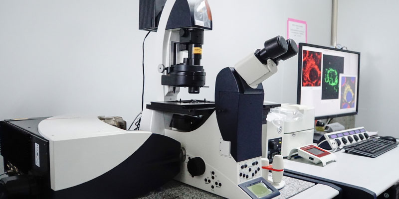
Confocal microscopy is an optical imaging technique that relies on spatial filtering methods to eliminate the contribution to the image from areas of the sample that are not immediately in focus.
1. Fundamentals of Confocal Microscopy
Confocal microscopy, the leader in fluorescence microscopy, enables fine imaging of cells and tissues by exciting different subgroups of fluorophores in the sample through laser technology. Unique to conventional epifluorescence microscopy is the high resolution when imaging thick samples, the effective elimination of out-of-focus glare caused by spatial filtering, and the significant reduction of photo-induced sample damage.
- Unique Featured Optics
Confocal microscopes use a laser to illuminate the sample, replacing the incoherent tungsten or mercury lamp light source of conventional microscopes. By scanning the laser across different focal points, the system is able to collect images at different depth planes (i.e. optical sections) of the sample. This optical sectioning technique makes it possible to image living samples without the need for physical sectioning, while also prolonging sample survival by reducing phototoxicity.
- Improvements in Image Performance
In terms of image performance, the Confocal Microscope System offers improved axial and lateral resolution compared to conventional fluorescence microscopes. In addition, its unique pinhole assembly design effectively blocks out-of-focus fluorescence to ensure image clarity and detail.
At the same time, confocal microscopy significantly optimises image contrast beyond conventional methods by reducing background noise and improving signal-to-noise ratio. With conventional techniques, fluorescence emitted from neighbouring parts of the sample interferes with the image plane, resulting in blurred focus, which makes it difficult to distinguish fluorescence that does not contribute to the imaging focal plane, especially in samples thicker than two microns. However, confocal microscopy utilises spatial filtering techniques to effectively solve this problem by blocking out-of-focus light that blurs the image. Although confocal and multiphoton microscopes have similar resolutions in the x- and y-axes, multiphoton microscopes are superior in the z-axis or depth resolution. Confocal microscopy uses only a single photon to excite the fluorescent dye, which increases light scattering in the image and limits the depth of imaging in the sample, but still provides better resolution than conventional wide-field techniques and serves as a bridge between classical wide-field techniques and more advanced super-resolution techniques. As a result, confocal microscopy excels in a wide range of applications.
2.Technical Details Explanation
- Important Components and Functions
In the field of microscopic imaging, confocal microscopy stands out for its outstanding optical performance. It is able to provide images that are finer and more focused than conventional microscopes, thus playing a crucial role in scientific research and medical diagnosis. Figure 2 depicts the typical construction of a confocal microscope, which contains four core components:
Laser excitation source: responsible for emitting laser light into the system for precise focusing on the sample.
Fluorescent dichroic filter: This filter reflects the excitation light back to the sample and allows the secondary fluorescence emitted by the sample to pass through the detection system.
Pinhole: A key component of the confocal unit, the pinhole acts as a spatial filter, effectively preventing non-objective focal plane light from interfering with the image.
Stepper motors: enable data collection in the x and y directions along the z-axis by controlling the point-by-point scanning of the laser beam across the sample. This helps to automatically collect 3D data, which in turn allows for easy construction of 3D images.
- Synergy of different components
Objective lenses: High numerical aperture (NA) objective lenses are essential for achieving high resolution. Water- or oil-immersion objective designs are often used to compensate for the difference in refractive index between the preservation medium for live samples and water. 3D imaging and optical sectioning techniques require long working distances.
optlenses
Related posts
Activity 11 Optics Of The Human Eye
What is The Surface Area of This Rectangular Prism Brainly?
How to Get Super Glue Off Eyeglass Lenses?
Why are Some Phone Companies Copying Iphone Camera Lenses?

