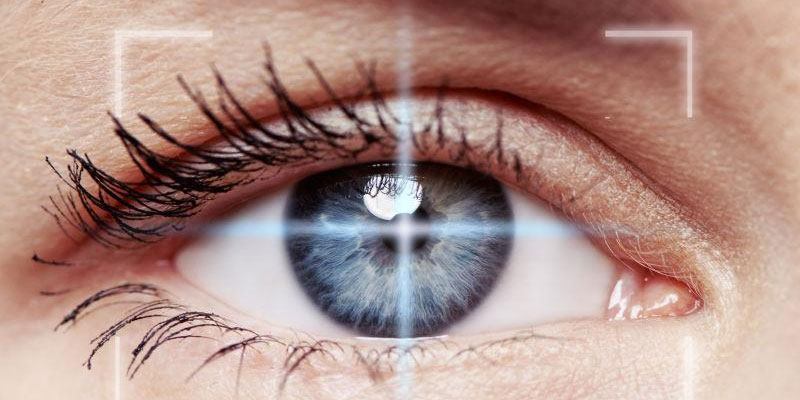
Activity 11 Optics Of The Human Eye is as follows: Light rays pass through the cornea and lens, where they are refracted and focused onto the retina. Photoreceptor cells convert these light signals into electrical signals, which are then transmitted via the optic nerve to the brain for processing into images. This entire process relies on optical refraction, bioelectric conduction, and neural coordination to ultimately form visual perception.
1.Light Entry and Focusing
- The refractive action of the cornea and lens Light first passes through the transparent cornea, undergoing its initial refraction. It then enters the lens, which further alters the angle of refraction by adjusting its shape (controlled by the ciliary muscle), enabling precise focusing of light onto the retina.
- Iris and Pupil Regulation The iris alters pupil size by contracting or dilating, controlling the amount of light entering the eye. Under bright light, the pupil constricts; under dim light, it expands—similar to a camera’s aperture mechanism.

2.Photoreception and Signal Conversion in the Retina
- Function of Photoreceptor Cells
Rod cells and cone cells in the retina are responsible for light sensitivity.
Rod cells: Sensitive to dim light (e.g., night vision) but unable to distinguish colors.
Cones: Responsible for perceiving bright light and color, concentrated in the macula at the center of the retina, providing high-resolution vision.
- Conversion of Light Signals into Electrical Signals
Light stimulates photopigments (e.g., rhodopsin) within photoreceptor cells, triggering chemical reactions that generate electrical signals. These signals are transmitted stepwise through bipolar cells and ganglion cells within the retina.
3.Transmission of Visual Signals and Brain Processing
- Conduction Function of the Optic Nerve
The axons of ganglion cells converge to form the optic nerve, transmitting electrical signals to the lateral geniculate nucleus (visual relay station) in the brain, which then projects to the visual cortex. - Visual Integration in the Brain
The brain fuses signals from both eyes, corrects the inverted retinal image, and integrates information such as color, shape, and motion to ultimately form a three-dimensional, coherent visual perception.
4.Auxiliary Structures and Protective Mechanisms of the Eye

- Sclera and Aqueous Humor
The sclera (white of the eye) maintains the shape of the eyeball; aqueous humor fills the anterior chamber, providing nutrients and maintaining intraocular pressure. - Tears and Eyelids
Tears lubricate the cornea and kill bacteria; eyelids remove foreign objects through blinking and evenly distribute tears. - Vitreous Humor
This transparent gel-like substance fills the posterior chamber of the eye, supporting the retina and maintaining the path for light transmission.
5.Scientific Explanations for Common Visual Phenomena
- Myopia and Hyperopia: Caused by an eyeball that is too long or too short, or abnormal lens accommodation, resulting in the focal point falling in front of or behind the retina.
- Color Blindness: A deficiency in a specific light-sensitive pigment within the cone cells, leading to impaired recognition of certain colors.
- Dark Adaptation: When moving from bright light into darkness, rod cells require several minutes to regain sensitivity, gradually enhancing night vision.
The functioning of the eye represents a perfect fusion of biological evolution and intricate physical structure, with each step relying on the coordinated action of specific tissues. Understanding its principles aids in scientifically protecting vision and recognizing the causes of visual impairments.
optlenses
Related posts
Activity 11 Optics Of The Human Eye
What is The Surface Area of This Rectangular Prism Brainly?
How to Get Super Glue Off Eyeglass Lenses?
Why are Some Phone Companies Copying Iphone Camera Lenses?


