Content:
Parts Of Microscope: the objective lens, eyepiece, light source, condenser, and auxiliary optical components.

1:Core optical components and functions
1.Objective Lens
- Position: Close to the specimen, mounted on the rotating objective turret.
- Function: Primary magnification, determining resolution and image quality. Objectives are typically labelled with magnification values such as ‘4×, 10×, 40×, 100×’. High-magnification objectives (e.g., 100×) require immersion oil for use.
- Structure: Multiple lens elements arranged in groups, with chromatic aberration correction (e.g., ‘plan achromatic objectives’) to enhance clarity.
2.Eyepiece/Ocular Lens
- Position: The top lens through which the observer’s eye makes contact.
- Function: Second magnification of the objective lens image, transmitting it to the human eye. Common magnifications are 10× or 15×. Total magnification = objective magnification × eyepiece magnification.
- Features: Some eyepieces incorporate built-in pointers or scales to assist in measuring or marking samples.
3.Illumination System
- Type: Traditional microscopes employ mirrors (reflecting natural light), whereas modern models predominantly utilise LED or halogen lamps as stable light sources.
- Function: Provides uniform illumination with adjustable brightness to accommodate different specimens (e.g., transparent/non-transparent).
4.Condenser
- Location: Below the stage, near the light source.
- Function: Focuses light onto the sample to enhance contrast and resolution.
- Adjustment: Features an aperture diaphragm (controls light intensity) and a vertical adjustment knob (adjusts the focal plane). Must be matched with the objective’s numerical aperture (NA value).
2:Auxiliary optical components:
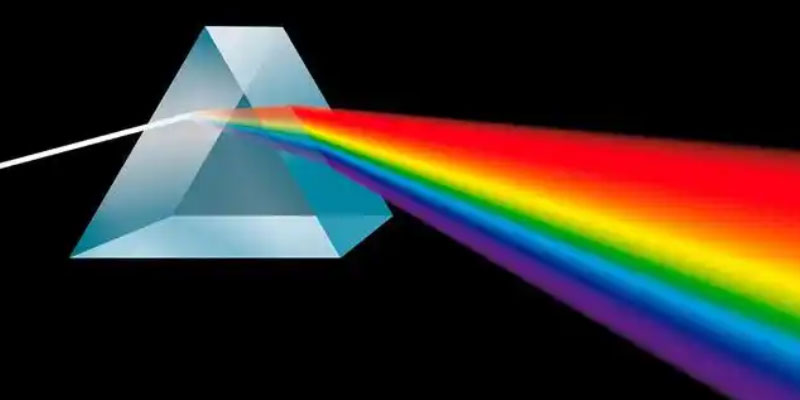
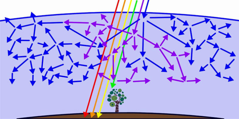
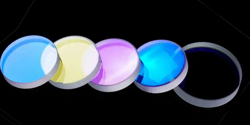
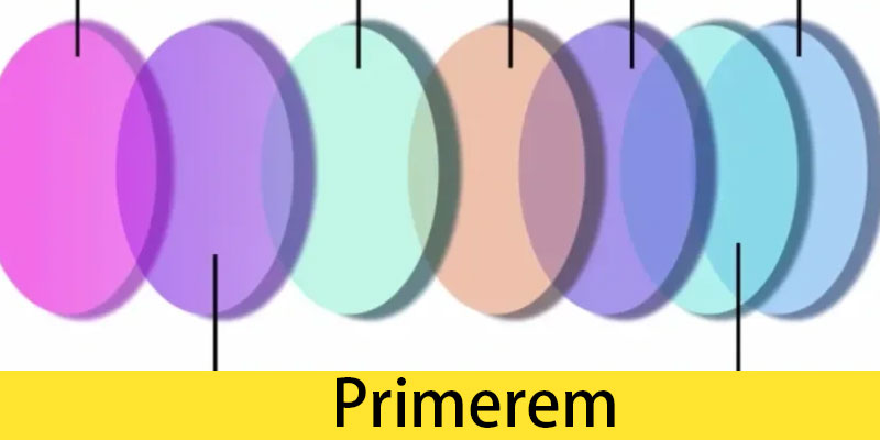
1.Filter
Function: Adjust light color (e.g., blue filters enhance contrast in white light) or block specific wavelengths (e.g., excitation filters in fluorescence microscopy).
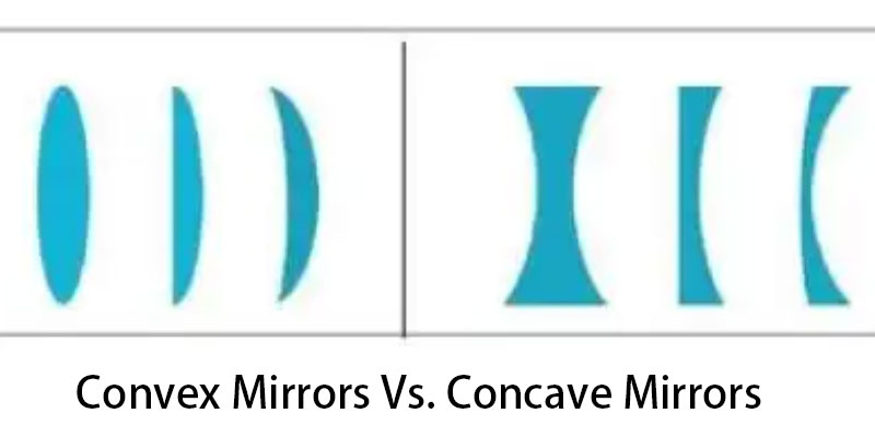
2.Mirror
Applicable scenarios: Microscopes without an internal light source, which reflect external light via plane/concave mirrors. Concave mirrors can gather more light.
3.Phase loop and differential interference element
Special purpose: For use in phase-contrast microscopy or differential interference contrast microscopy (DIC), enhancing contrast in transparent specimens.
3:The principle of collaborative operation of the optical system:
- Optical path: Light source → Condenser → Specimen → Objective lens → Eyepiece → Human eye.
- Key adjustments:
Köhler illumination: Align the condenser with the light source to ensure uniform illumination across the specimen.
Aperture matching: The condenser aperture must correspond to the numerical aperture of the objective lens to prevent halos or reduced resolution.
4:Maintenance and Usage Tips:
- Cleaning: Wipe optical components with lens paper and specialised cleaning solution to avoid scratching the coating.
- Dust protection: Cover with a dust cap after use to prevent dust entering the optical path.
- Moisture protection: Store in a dry environment to prevent mould growth on the lenses.
By understanding the functions and cooperative relationships of these optical components, one can operate the microscope more efficiently and obtain high-quality images.
optlenses
Related Blogs